Translate this page into:
Exploring the kaleidoscope: Ocular manifestations in leukaemia – A case series revelation
*Corresponding author: Krutika Kamalakar Thorat, Department of Ophthalmology, MGM Medical College and M Y Hospital, Indore, Madhya Pradesh, India. krutikathorat99@gmail.com
-
Received: ,
Accepted: ,
How to cite this article: Walia S, Thorat KK. Exploring the kaleidoscope: Ocular manifestations in leukaemia – A case series revelation. Glob J Cataract Surg Res Ophthalmol. 2024;3:108-12. doi: 10.25259/GJCSRO_19_2024
Abstract
Leukaemia is a systemic cancer of the blood and blood-forming tissues that affects multiple organs. Acute lymphocytic leukemia (ALL) is the dominant leukaemia type in children and more than 50% of these patients can be cured. In adults, acute myelocytic leukemia (AML) is the predominant myeloproliferative disorder, with lower survival rates. Around 35.4% of leukaemia patients present with leukaemic retinopathy. The purpose of the study is to present ocular manifestations in acute leukaemia. This report outlines the clinical presentations of three patients. Among them, one adult presented with AML at diagnosis, exhibiting ocular symptoms. Another adult, 6 months after diagnosis, experienced retrobulbar haemorrhage and leukaemic retinopathy. In addition, a paediatric patient, previously treated for ALL and in remission, developed unilateral vision loss, subsequently indicating disease relapse. During the diagnosis of acute leukaemia patients, it is recommended to conduct a thorough ophthalmic evaluation, which should include a dilated fundus examination, as ocular involvement in these patients is prevalent and may occasionally be asymptomatic.
Keywords
AML
Retinal haemorrhage
Optic nerve infiltration
Leukaemic retinopathy
Intraretinal haemorrhage
INTRODUCTION
Leukaemias, malignancies originating from haematopoietic stem cells, are categorised based on clinical presentation. Acute leukaemias exhibit rapid onset and accompanying symptoms such as anaemia, haemorrhage and infection, while chronic leukaemias typically manifest initially with vague symptoms in an indolent manner. Ophthalmic manifestations of leukaemia are evident in both acute and chronic forms of the disease.
Leukaemic ophthalmopathy can be classified into primary infiltration, where leukaemic cells directly invade eye tissues, and secondary lesions resulting from factors such as increased blood thickness or compromised immunity. Acute lymphocytic leukemia (ALL) is the dominant leukemia type in children while in adults, acute myelocytic leukemia (AML) is the predominant myeloproliferative disorder.[1] Patients may either be asymptomatic or experience vision impairment.[2] It is noted that approximately 35.4% of leukaemia patients display leukaemic retinopathy.[3]
CASE SERIES
Case 1
A 30-year-old man presented to M Y Hospital, Indore, with the chief complaints of fever, nosebleed and difficulty in swallowing. In addition, he developed a diminution of vision in both eyes over the past 20 days. Snellen visual acuities recorded visual acuity in both eyes by counting fingers at 1 foot with accurate projection of rays.
Pupillary examination showed normal pupils with brisk reactions to light and no relative afferent pupillary defect (RAPD). Anterior segment examination indicated normal findings, with clear cornea and deep, quiet anterior chambers in both eyes, along with a normal iris in both eyes.
A dilated fundus examination was conducted using 1% tropicamide. The lens and vitreous humour of the right eye (RE) appeared clear. A cup-to-disc ratio (CDR) of 0.2 was noted, alongside dot and blot haemorrhages and intraretinal haemorrhages in all quadrants covering the posterior pole and fovea of the RE. Roth’s spots and cotton wool spots were also observed. The absence of foveal reflex was noted, along with venous dilation in all quadrants of the fundus [Figure 1].

- Case 1-Photograph Showing multiple Roth’s Spot, cotton wool spots, hemorrhages. RE: Right eye, LE: Left eye.
In the left eye (LE), the CDR was not appreciable, and a similar haemorrhagic fundal presentation was observed, although with more haemorrhages concentrated in the posterior pole.
At this stage, a full blood count with differential at the haematology visit revealed a white blood cell count of 105.42 × 10³/μL, along with a neutrophil count of 53.03 × 10³/μL, a lymphocyte count of 40.27 × 10³/μL and a mid-granulocyte count of 11.50 × 10³/μL. The haematocrit level was 21%, mean corpuscular volume (MCV) was 87.20, mean corpuscular haemoglobin (MCH) was 37.60 and mean corpuscular haemoglobin concentration (MCHC) was 43.20, indicative of anaemia. The platelet count was 35.00 × 10³/μL, indicative of thrombocytopenia. These features were suggestive of acute leukaemia.
The patient underwent a bone marrow biopsy, which showed granulopoiesis (active, dominated by promyelocytes in 50% of the nucleated marrow cells), and he was diagnosed with acute promyelocytic leukaemia (PML). Fluorescence in situ hybridisation for PML/retinoic acid receptor alpha was ordered and came back negative.
The patient was promptly initiated on treatment with alltrans retinoic acid[4] and hydroxyurea. They were advised to return for follow-up care and ophthalmological monitoring of macular haemorrhage. After 2 months of hydroxyurea therapy, a repeat complete blood count showed improved results: White cell count of 1.78 × 10⁹/L, neutrophil count of 0.69 × 10⁹/L, lymphocyte count of 0.55 × 10⁹/L and monocyte count of 0.11 × 10⁹/L, indicating a favourable response to treatment. The persistence of anaemia, reflected by a red blood cell count of 3.23 × 10¹²/L and a haematocrit level of 28.9%, was expected with hydroxyurea therapy. After the initial treatment, oral imatinib tablets (400 mg/day) were administered.
After treatment, intraretinal haemorrhages and dot blot haemorrhages in both eyes decreased, and cotton wool spots also diminished. The LE regained its foveal reflex, leading to improved visual acuity of 6/60 in the LE and 6/24 in the RE.
Case 2
An 8-year-old male presented to M Y Hospital, Indore, with the chief complaint of the sudden loss of vision in the LE over the past 15 days. The onset was abrupt, painless and not associated with any other ocular complaints. Snellen visual acuity assessment revealed visual acuity in the RE as 6/9 (p), improving to 6/6, while in the LE, there was no perception of light. Pupillary reaction in the LE showed a RAPD. Slit-lamp assessment of the anterior segment revealed normal findings.
The patient had a known history of acute T-cell lymphocytic, for which he underwent treatment including vincristine, cytarabine, bone marrow transplant, intrathecal chemotherapy for central nervous system (CNS) prophylaxis and is currently receiving methotrexate and 6-mercaptopurine.
A dilated fundus examination was conducted using a drop of 1% tropicamide. The lens and vitreous humour of the RE appeared clear, with a CDR of 0.2 and a present foveal reflex. The general fundus exhibited normal characteristics. In the LE, the media was clear, but the disc showed blurred margins in all quadrants, with an appreciable CDR. Vascular dilatation and sheathing were observed in the blood vessels, along with pigmentary changes at the macula. The remainder of the fundus appeared normal, indicative of possible optic nerve infiltration [Figure 2].

- Case 2 - Left eye fundus showing blurred disc margin with unappreciable cup disc ratio. RE: Right eye LE: Left eye.
The patient, who was suspected to have experienced a relapse, was referred to a haematologist-oncologist for additional management.
Case 3
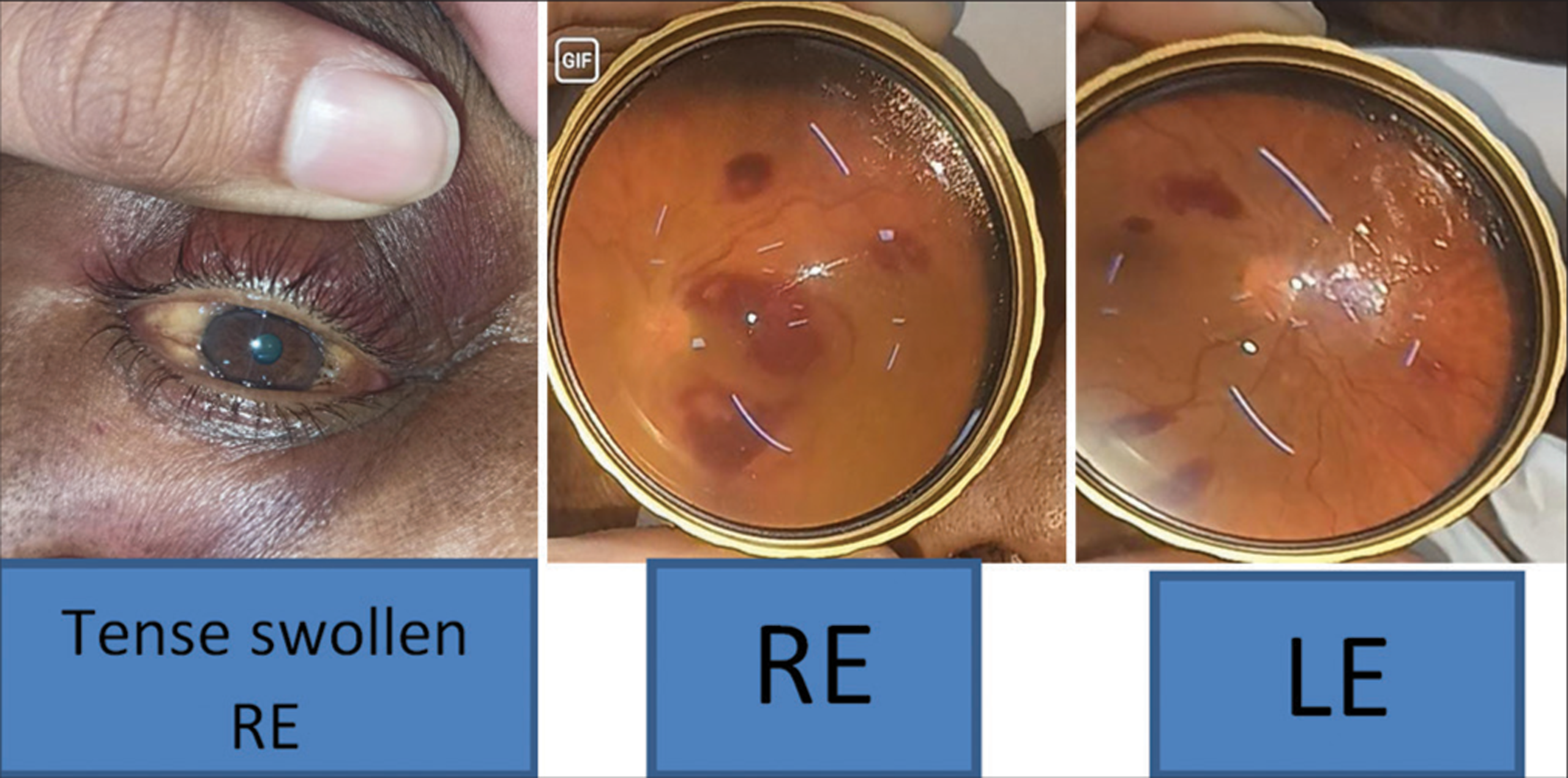
A 50-year-old male presented to M Y Hospital, Indore, with the chief complaint of swelling in the RE, accompanied by pain and mild watering for the past 2 days, without any history of trauma. Snellen visual acuity assessment revealed visual acuity in the RE as 5/60, not improving with pinhole, and in the LE as 6/36, improving with pinhole to 6/18. Anterior segment examination of the RE showed oedematous lids, chemosed conjunctiva and restricted extraocular movements in all cardinal gazes. The pupillary reaction was central and circular, reacting to light, while the LE appeared normal.
The patient had a known history of AML for the past 6 months and was undergoing cycles of cytarabine and doxorubicin but was non-compliant with medication.
A dilated fundus examination was performed using a drop of 1% tropicamide. The lens and vitreous humour of the RE appeared clear. Blurred disc margins were observed, and the CDR was not appreciable. Blood vessels exhibited dilation, tortuosity and perivascular sheathing. Multiple subretinal haemorrhages were present in both the macular and midperipheral areas, along with Roth’s spots.
In the LE, media clarity was noted, with a distinct circular disc margin and a CDR of 0.3:1. Dilated blood vessels, vascular sheathing and a present foveal reflex were observed. Multiple subretinal haemorrhages were also noted in the midperipheral retina [Figure 3].

- Case 3 - Right eye is tense swollen Fundus examination of right eye showing multiple subretinal hemorrhages, with tortuous blood vessels and blurred disc margins. Fundus examination of the left eye shows multiple subretinal hemorrhages with tortuous blood vessels. RE: Right Eye, LE: Left Eye.
A full blood count with differential during the haematology visit revealed a white blood cell count of 2.33 × 10³/μL, with a neutrophil count of 0.32 × 10³/μL (13.9%), and a lymphocyte count of 1.77 × 10³/μL (75.9%), suggestive of leukopenia and granulopenia. Haematocrit level was measured at 10.5%, with MCV at 77.4, MCH at 22.5 and MCHC at 29, indicating anaemia. Thrombocytopenia was evident with a platelet count of 19.00 × 10³/μL. Serum ferritin levels were elevated at 1079.00 ng/mL, while total iron-binding capacity was 150.70 and unsaturated iron binding capacity was 81.20 μg/dL. Serum creatinine level was 0.45 mg/dL, and blood glucose level was 104.8 mg/dL. These findings collectively suggest acute myeloid leukaemia.
On ultrasonography B-scan, retrobulbar haemorrhage and compartment syndrome of the RE was detected. As a result, a lateral canthotomy was performed. The patient was expeditiously referred to a haematologist-oncologist for comprehensive management of AML. Follow-up appointments were arranged by evaluating the treatment response, with the next consultation scheduled after a 2-month interval. Following appropriate intervention, visual acuity in the RE improved to 6/36, while the LE achieved a visual acuity of 6/12.
DISCUSSION
The ocular manifestations of leukaemic retinopathy are diverse. Previously reported findings have included flame-shaped haemorrhages, cotton wool spots, Roth spots (white-centred haemorrhages), retinal venous tortuosity and neovascularisation.[5] Leukaemic retinopathy is commonly seen at the posterior pole or may extend into vitreous or subretinal spaces.[6]
Occasionally, the ophthalmic symptoms and findings can be the initial manifestation of the systemic illness or may present as a disease relapse.[7]
The occurrence of ocular manifestations at the time of diagnosis in the first case is notably uncommon, as corroborated by existing literature. The presence of bilateral intraretinal haemorrhages, Roth’s spots and cotton wool spots underscores the pivotal role of ocular examination in the initial diagnostic assessment of patients, particularly those with systemic conditions such as AML.
Optic nerve infiltration may be the first presenting sign of acute leukaemic relapse before the haematological involvement.[8,9] The optic nerve is known to be a sanctuary for leukaemic cells which are relatively unaffected by systemic chemotherapy and even intrathecal chemotherapy.[9] Cases of both unilateral and bilateral optic nerve involvement have been reported.[10,11]
In our case, despite intrathecal chemotherapy, which was given for CNS prophylaxis at the first diagnosis of ALL and achieving complete remission after completion of chemotherapy, the case developed isolated CNS relapse.
Furthermore, the relapse presenting as optic nerve infiltration highlights the aggressive nature of T-acute lymphocytic leukaemia (T-ALL) and the propensity for extramedullary spread, despite previous treatment regimens. This underscores the importance of vigilant surveillance for disease recurrence, even in cases where initial treatment achieves remission.
In the last case, the patient exhibited retrobulbar haemorrhage, a rare yet serious complication of leukaemia, requiring immediate intervention through lateral canthotomy. This urgent procedure aimed to relieve compartment syndrome and safeguard visual function. The subsequent diagnosis of AML highlights the significance of contemplating systemic malignancies in patients with ocular manifestations, especially when concurrent with haematologic irregularities.
The improvement in visual acuity post-treatment emphasises the possibility of recovery with timely and suitable intervention. Nevertheless, the noted enhancement to 6/36 in the affected eye and 6/12 in the opposite eye accentuates the lasting influence of leukaemic retinopathy on visual function, persisting even after intervention.
The treatment for leukaemic retinopathy does not involve any direct therapy. Chemotherapy, immunotherapy and radiotherapy, however, are usually implemented to manage the underlying systemic cause.[1] In 2018, Lyu et al. suggested that expectant management should be the first-line treatment of choice due to the higher risk of bleeding and infection in leukaemic patients.[12]
A study conducted by Abu El-Asrar et al. revealed that patients presenting with leukaemic retinopathy exhibited a poorer prognosis compared to those without such ocular manifestations. This observation underscores the potential prognostic significance of leukaemic retinopathy in the context of systemic leukaemia.[13]
Similarly, Ohkoshi and Tsiaras evaluated the prognostic implications of leukaemic retinopathy in childhood leukaemia among 63 patients. Their findings indicated a notable difference in the 5-year survival rate between patients with leukaemic retinopathy at presentation and those without ophthalmic involvement. Specifically, individuals with leukaemic retinopathy exhibited a significantly lower 5-year survival rate compared to their counterparts without ocular manifestations, with rates of 21.4% and 45.7%, respectively.[14]
Gross leukaemic infiltrates, as illustrated, have been identified in moribund patients.
CONCLUSION
Retinal haemorrhage is a serious complication in leukaemic patients; it could be the presenting complaint or it may even manifest after the initiation of therapy. It could result from direct leukaemic pathological infiltrates or secondary to anaemia, thrombocytopenia or hyperviscosity.
Relapse can be in the form of optic nerve infiltration that can occur despite prophylactic CNS chemotherapy or radiotherapy.
Timely detection and regular follow-ups are imperative for effective management. These cases highlight the importance of routine eye examinations that ophthalmologists should perform in leukaemic patients.
Ethical approval
Institutional Review Board approval is not required.
Declaration of patient consent
The authors certify that they have obtained all appropriate patient consent.
Conflicts of interest
There are no conflicts of interest.
Use of artificial intelligence (AI)-assisted technology for manuscript preparation
The authors confirm that there was no use of artificial intelligence (AI)-assisted technology for assisting in the writing or editing of the manuscript and no images were manipulated using AI.
Financial support and sponsorship
Nil.
References
- A case of leukemic retinopathy mimicking common ischemic retinopathies. Clin Case Rep. 2015;4:133-7.
- [CrossRef] [PubMed] [Google Scholar]
- Ocular and orbital involvement in leukemia. Surv Ophthalmol. 1983;27:211-32.
- [CrossRef] [PubMed] [Google Scholar]
- Use of all-trans retinoic acid in the treatment of acute promyelocytic leukemia. Blood. 1988;72:567-72.
- [CrossRef] [PubMed] [Google Scholar]
- Retinopathy in acute leukaemia at initial diagnosis: Correlation of fundus lesions and haematological parameters. Acta Ophthalmol Scand. 2004;82:81-5.
- [CrossRef] [PubMed] [Google Scholar]
- Ophthalmic manifestations of leukemia. Curr Opin Ophthalmol. 2016;27:545-51.
- [CrossRef] [PubMed] [Google Scholar]
- Chronic myeloid leukemia presenting with bilateral central retinal vein occlusion and massive retinal infiltrates. J AAPOS. 2012;16:406-8.
- [CrossRef] [PubMed] [Google Scholar]
- Leukaemic infiltration of the optic nerve as the initial manifestation of leukaemic relapse. Eye. 2004;18:546-50.
- [CrossRef] [PubMed] [Google Scholar]
- Bilateral central retinal artery occlusion associated with leukemic optic neuropathy. Can J Ophthalmol. 2007;42:759-60.
- [CrossRef] [PubMed] [Google Scholar]
- Leukemic involvement of the optic nerve. Am J Ophthalmol. 1988;105:294-8.
- [CrossRef] [PubMed] [Google Scholar]
- Acute bilateral retina hemorrhages beneath internal limiting membrane: An unusual ophthalmological case report of acute leukemia during complete clinical remission. Medicine. 2018;97:e0000.
- [CrossRef] [PubMed] [Google Scholar]
- Prognostic importance of retinopathy in acute leukemia. Doc Ophthalmol. 1995;91:273-81.
- [CrossRef] [PubMed] [Google Scholar]
- Prognostic importance of ophthalmic manifestations in childhood leukaemia. Br J Ophthalmol. 1992;76:651-5.
- [CrossRef] [PubMed] [Google Scholar]


